Myopia Management
Slows Myopia Progression |Reduce Risk of Serious Eye Conditions|Improve Quality of Life
Myopia, commonly known as nearsightedness, is a vision condition where close objects appear clear, but distant objects are blurry. This occurs when the eye grows too long from front to back or the cornea becomes too curved, causing light to focus in front of the retina instead of directly on it.
Myopia control therapy includes various treatments and strategies designed to slow the progression of myopia in children and young adults. The goal is to reduce the risk of developing severe myopia, which is associated with a higher risk of serious eye problems such as retinal detachment, glaucoma, and cataracts later in life.
If your child is showing signs of myopia, early intervention is crucial. Schedule a consultation with Dr. Priya to discuss myopia control therapy and find the best treatment plan for your child. Our experienced and compassionate team is dedicated to providing exceptional care and helping you protect your child's vision for the future.
Take the first step towards better vision and a brighter future for your child!
 Request An Appointment

5 options of
Myopia Control Therapy
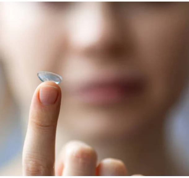
| 01 | Orthokeratology (Ortho-K) | Specially designed gas-permeable contact lenses are worn overnight to temporarily reshape the cornea, providing clear vision during the day without the need for glasses or contact lenses. |
|---|---|---|
| 02 | Atropine Eye Drops | Low-dose atropine eye drops are used to slow the progression of myopia. Studies have shown that atropine can be effective in reducing the rate of eye growth in myopic children. |
| 03 | Multifocal Contact Lenses | These contact lenses are designed with multiple zones for different vision distances. They help to reduce the strain on the eyes and slow the progression of myopia. |
| 04 | Multifocal Eyeglasses | Similar to multifocal contact lenses, these glasses have different lens powers to correct vision at various distances, helping to control myopia progression. |
| 05 | Lifestyle and Environmental Modifications | Encouraging outdoor activities and reducing prolonged near-work tasks like reading or screen time can help slow myopia progression. |